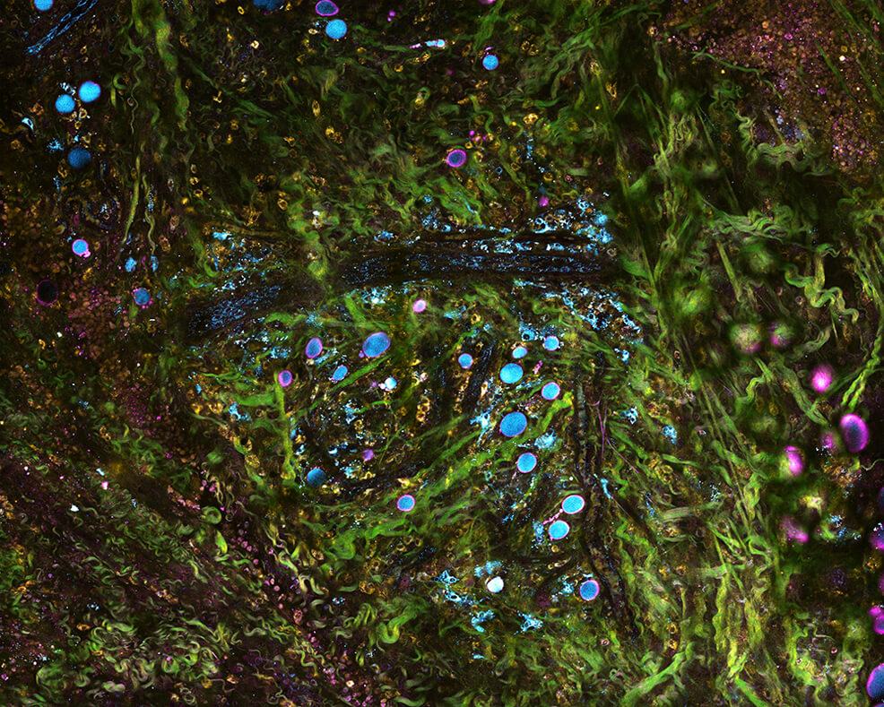
22nd June 2018 New tissue-imaging technology could enable real-time diagnostics A new microscope system can image living tissue in real time and in molecular detail, without any chemicals or dyes, report researchers at the University of Illinois. The system uses precisely tailored pulses of light to simultaneously image with multiple wavelengths. This enables the researchers to study concurrent processes within cells and tissue, and could give cancer researchers a new tool for tracking tumour progression and physicians new technology for tissue pathology and diagnostics. The researchers describe their technique – called simultaneous label-free autofluorescence multi-harmonic microscopy (SLAM) – in the peer-reviewed journal Nature Communications.
"The way we have been removing, processing and staining tissue for diagnosing diseases has been practiced the same way for over a century," said study leader Dr. Stephen Boppart, a professor of bioengineering at Illinois and a medical doctor. "With advances in microscopy techniques such as ours, we hope to change the way we detect, visualise and monitor diseases that will lead to better diagnosis, treatments and outcomes." SLAM microscopy differs from standard tissue pathology in several ways. First, it is used on living tissue, even inside a living being, giving it the potential to be used for clinical diagnosis or to guide surgery in the operating room. Second, it uses no dyes or chemicals, only light. Standard procedure involves removing a tissue sample and adding chemical stains – which can be a lengthy process – and the chemicals can disrupt the cells. While other stain-free imaging techniques have been developed, they usually only visualise a subset of signals, measuring specific biological or metabolic signatures, explains graduate student Sixian You, the first author of the paper. Meanwhile, SLAM microscopy simultaneously collects multiple contrasts from cells and tissues, capturing molecular-level details and dynamics such as metabolism. In their most recent study, Boppart's group looked at mammary tumours in rats, along with the surrounding tissue environment. Thanks to the simultaneous data, they were able to observe the range of dynamics as the tumours progressed and how different processes interacted. "We know the tumour is there, but tumours support a whole ecosystem in the tissue," said You. "They recruit healthy cells to support them. SLAM allows us to have a comprehensive picture of this ever-evolving tumour micro-environment at subcellular, molecular and metabolic levels in living animals and human tissue. Monitoring that process can help us better understand cancer progression – and in the future, could lead to better diagnosis of how advanced a tumour is, and better therapeutic approaches aimed at halting the progression." In the video above, SLAM microscopy follows a white blood cell (called a leukocyte) as it travels through a blood vessel. "There is a wealth of new data, information and biomarkers in the images we collect from fresh tissue that is still metabolically active, or in living organisms, where we can visualise the dynamics of individual cells and their collective behaviours," explains Boppart. "These, we expect, will become new markers for diseases such as cancer – and our imaging technology will help detect these for disease screening, diagnostics and monitoring applications. We believe that this technology will open the possibility of complementing, or even replacing, standard histopathology processing, which is time-and labour-intensive."
Comments »
If you enjoyed this article, please consider sharing it:
|