24th October 2020 World record resolution in cryo-electron microscopy A novel technique developed by Max Planck researchers in Germany can visualise individual atoms in a protein with cryo-electron microscopy for the first time.
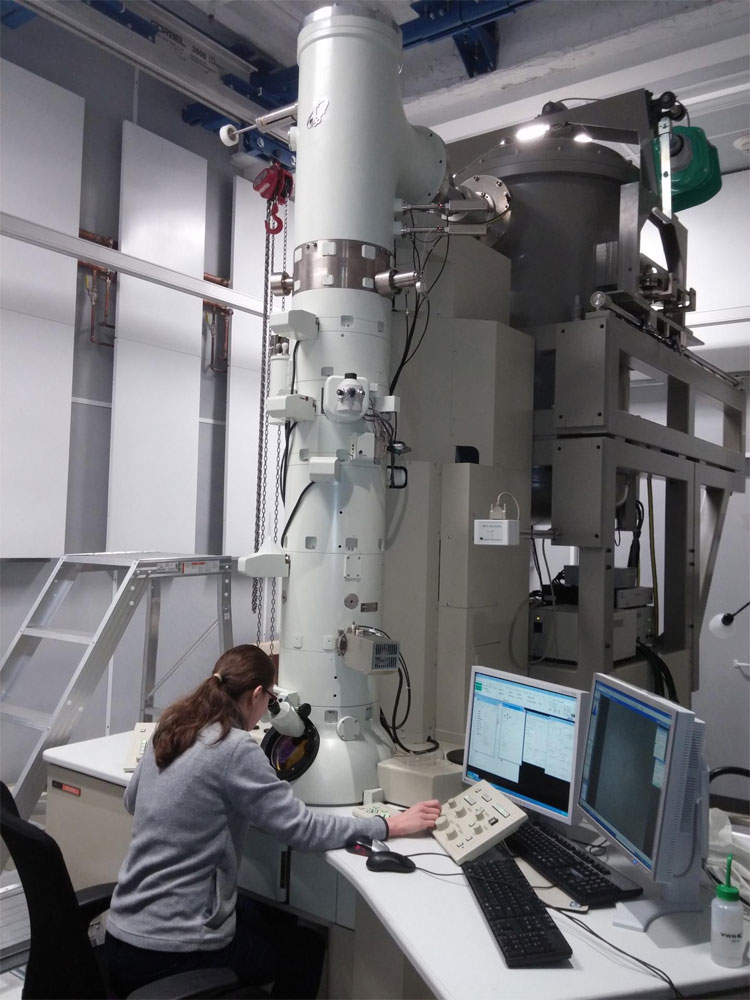
A crucial resolution barrier in cryo-electron microscopy has been broken. Scientists at the Max Planck Institute (MPI) for Biophysical Chemistry have observed single atoms in a protein structure for the first time, and taken the sharpest ever images using this method. Such unprecedented details are essential to understand how proteins function in cells or cause diseases. The technique could also be used to develop active compounds for new drugs in the future. Since the outbreak of the COVID-19 pandemic, scientists around the world have been working to determine the 3D structures of important key proteins of the novel coronavirus. Their common goal is to find docking sites for an active compound able to combat the pathogen effectively. One method for determining these molecular structures is cryo-electron microscopy (cryo-EM), a state-of-the-art technology whose developers recently won the Nobel Prize in Chemistry. The process involves quickly "freezing" a molecule in place and then bombarding it with electrons to make thousands of 2D projections, which are stacked in layers and combined to create a 3D reconstruction.
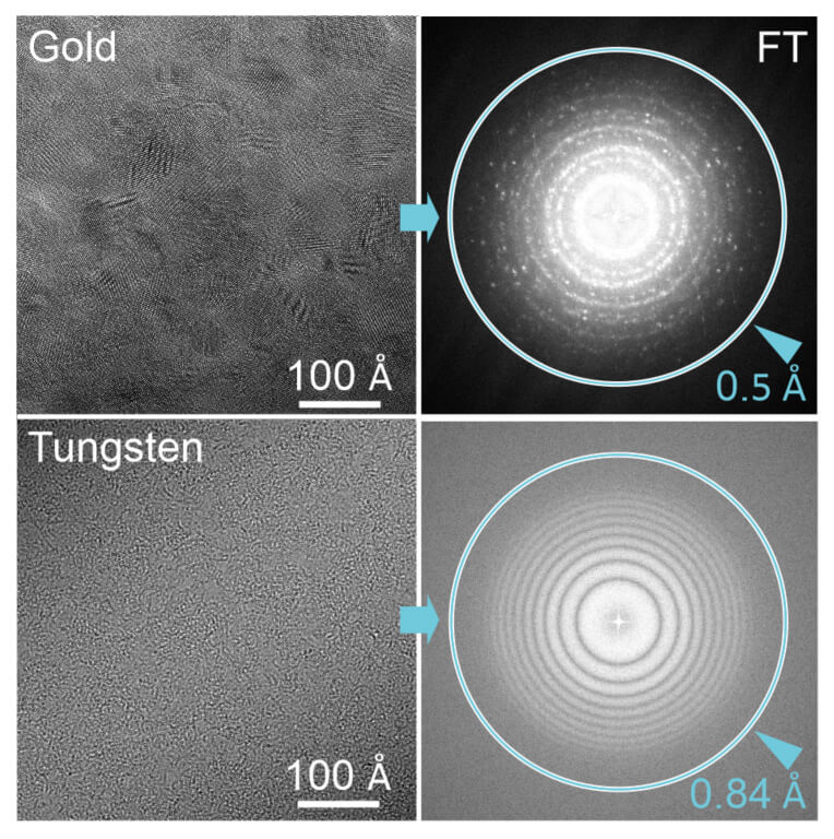
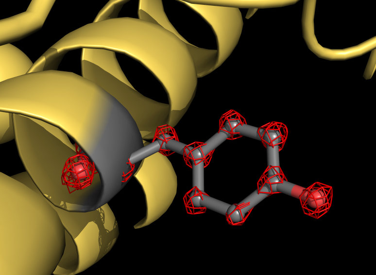
Holger Stark is a Professor in the Department of Structural Dynamics at the MPI. He and his team recently broke the world record for cryo-EM resolution, with a unique microscope setup they developed. "We equipped our device with two additional electron-optical elements to further improve image quality and resolution," explains Stark. "These ensure that imaging errors of optical lenses, so-called aberrations, no longer play a role." His doctoral student, Ka Man Yip, adds: "Electron microscopes are optical instruments and physically resemble a camera. The aberrations of an electron microscope interfere in cryo-EM in much the same way as those of a camera in photography. For a much-improved image quality it was therefore crucial to avoid these aberration errors." The new microscope allowed the scientists to capture more than one million images of a protein called apoferritin – mapping its molecular structure with a resolution of 1.25 angstroms. One angstrom is just 0.1 nanometres (nm), or a ten millionth of a millimetre. "We can now visualise single atoms in the protein – a milestone in our field," explains Stark. "For us, it was like putting super glasses on the microscope. The new structure reveals details never seen before. We can even see the density for hydrogen atoms and single atom chemical modifications."
To understand the inner workings of a human-made machine like a car engine, space rocket, or the cogs inside a clock, one must observe its different components directly at work. The same is also true for proteins, which are the "nanomachines" of natural, living cells. To get an idea of how they carry out tasks, one must know the exact position of all atoms of the protein. The ability to observe protein structures in such unprecedented detail will therefore bring major benefits for the world of medicine – accelerating structure-based drug design, for example, with new compounds tailored in the best and most accurate way for binding to viral proteins and blocking their function. Knowledge of the underlying mechanism of inhibition, and seeing how a compound and viral protein interact at the atomic level, could provide novel insights that help to produce more effective drugs with fewer side effects. "With breaking this cryo-EM resolution barrier, the technique has now reached a level where the benefits for pharmaceutical developments are directly visible," concludes Stark. It is now conceivable that cryo-EM will reach even subatomic resolutions in the future, says the team. A nearly ten-fold improvement in the highest resolution has already been achieved since the year 2000. Further orders of magnitude improvements therefore seem possible in the next few decades.
--- Follow us: Twitter | Facebook | Instagram | YouTube
Comments »
If you enjoyed this article, please consider sharing it:
|