7th October 2021 1 million neurons tracked in real time Scientists at Rockefeller University have developed a new technique for visualising the activity of a million neurons, near-simultaneously and with crystal clear resolution.
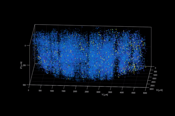
Until now, two-photon scanning microscopy combined with fluorescent tags has been the gold standard for imaging the activity of neurons within less transparent brain tissues, which are prone to scattering light. This involves firing a focused laser pulse at a tagged target. A few nanoseconds after the pulse hits, the tag emits fluorescent light that can show the approximate level of neuroactivity detected. However, two-photon microscopy suffers from a fundamental limitation. To understand the brain more fully, neurobiologists need to record simultaneous interactions between the sensory, motor, and visual regions – but capturing this activity across such a broad swathe of the brain is difficult without sacrificing resolution or speed. A new method – which has been dubbed "light beads microscopy" – is described in the journal Nature. This offers a creative solution that pushes the limits of imaging speed and is limited only by the physical nature of fluorescence itself. It eliminates the "deadtime" between sequential laser pulses when no neuroactivity is recorded and at the same time the need for scanning. The technique breaks one strong pulse into 30 smaller sub-pulses, each at a different strength, which dive into 30 different depths of scattering but induce the same amount of fluorescence at each depth. This is accomplished with a cavity of mirrors that staggers the firing of each pulse in time and ensures that they can all reach their target depths via a single microscope focusing lens. Using this approach, the only limit to the rate at which samples can be recorded is the time it takes the fluorescent tags to flare. That means broad swathes of the brain can be recorded within the same time it would take a conventional two-photon microscope to capture a much smaller network of brain cells. Scientists at Rockefeller University, New York, integrated their new system into a microscopy platform with access to a large brain volume. This enabled the recording of activity in more than a million neurons across the entire cortex of a mouse brain for the first time. "Understanding the nature of the brain's densely interconnected network requires developing novel imaging techniques that can capture the activity of neurons across vastly separated brain regions at high speed and single-cell resolution," said Alipasha Vaziri, PhD, Professor of Neurosciences and Behaviour at Rockefeller. "Light beads microscopy will allow us to investigate biological questions in a way that had not been possible before." A decade ago, scientists had been limited to scanning a few thousand neurons in real time. The orders of magnitude improvement since then suggests it may be possible to observe a whole mouse brain (70 million neurons) in the near future. Larger brains, such as those of monkeys (6.3 billion for a macaque) could follow – and then perhaps humans (86 billion) a relatively short time after that. In the coming decades, researchers might achieve "thought mapping" and psychological diagnosis at a near-real-time speed, making the treatment of both physical and mental illnesses much more targeted.
Comments »
If you enjoyed this article, please consider sharing it:
|