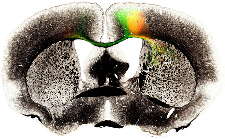
Neuroscientists reach major milestone in whole-brain circuit mapping 2nd June 2012 Neuroscientists at Cold Spring Harbor Laboratory (CSHL) reached a major milestone this week – publicly releasing the first data from their groundbreaking project, to construct the first whole-brain wiring diagram of a vertebrate brain, that of the mouse. The data, which totals 500 terabytes, consists of gigapixel images (a billion pixels each) of whole-brain sections. These can be zoomed to show individual neurons and their processes, creating a "virtual microscope." The images are integrated with other data sources from the web, and are being made fully accessible to neuroscientists as well as interested members of the general public (http://mouse.brainarchitecture.org). The data are being released pre-publication in the spirit of open science initiatives that have become familiar in digital astronomy (e.g. Sloan Digital Sky Survey) but are not yet as widespread in neurobiology.
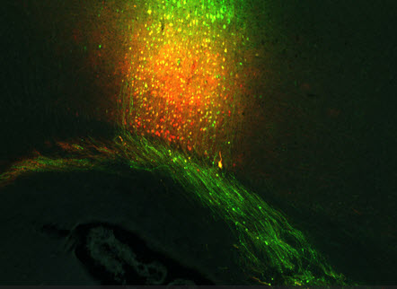
Each sampled brain is represented in about 500 images, each image being an optical section through a 20 micron-thick slice of tissue. Users can journey through each brain from "front" to "back", following the pathways taken through 3D brain space by tracer-labeled neuronal pathways. The tracers were picked to follow neuronal inputs and outputs of different brain regions. "Our project seeks to address a remarkable gap in our knowledge of the brain," says Partha Mitra, Professor of Biomathematics at CSHL and director of the Mouse Brain Architecture (MBA) Project. "Our knowledge of how the brain is wired remains piecemeal and partial after a century of activity. To understand how the brain works (or fails to work in neurological disease), it is critical that we understand this wiring diagram more fully. Further, there remain fundamental questions about brain evolution that cannot be addressed without obtaining such wiring diagrams for the brains of different species."
The MBA Project is distinguished by the approach advocated by Mitra in a 2009 paper. Back then, Mitra proposed mapping vertebrate brains at what he described as the “mesoscopic” scale – a mid-range amenable to light microscopy, giving far more detail than MRI-based methods, yet considerably less detail than via electron microscopy (EM). The latter, while useful for mapping synaptic connections between individual neurons, is feasible on a whole-brain basis only for tiny brains (e.g. fruitflies) or very small portions of the mouse brain. The pragmatic approach Mitra advocated, and which is realised in this first data release, is to image whole mouse brains in a semi-automated, quality-controlled process using light microscopy and injected neural tracers. While the basic methodology has been available for some time, systematically applying it to a grid of locations spanning the entire brain, and digitising/re-assembling the resulting collection of brains, is a new approach made feasible by the exponentially falling costs of computer storage. A mouse brain, at light-microscope resolution, generates around one terabyte (1000 GB) of data. Thus, producing and storing the data sets currently being gathered would have been impossibly expensive a decade or so ago. “Our project is what I’d call a necessary first step in a much larger enterprise – that of understanding both structure and dynamics of the vertebrate, and ultimately, the human brain,” says Mitra. Project highlights: http://brainarchitecture.org/mouse/highlights
Comments »
|