27th February 2014 Single chip device to provide real-time 3-D images from inside the heart and blood vessels Researchers have developed the technology for a catheter-based device that would provide forward-looking, real-time, 3-D imaging from inside the heart, coronary arteries and peripheral blood vessels. With its volumetric imaging, the new device could better guide surgeons working in the heart, and potentially allow more of patients' clogged arteries to be cleared without major surgery.
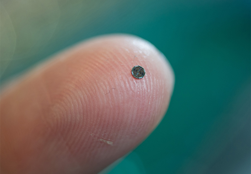
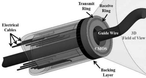
The device integrates ultrasound transducers with processing electronics on a single 1.4 millimetre silicon chip. On-chip processing of signals allows data from more than 100 elements on the device to be transmitted using just 13 tiny cables, permitting it to easily travel through circuitous blood vessels. The forward-looking images produced by the device would provide significantly more information than existing cross-sectional ultrasound. Researchers have developed and tested a prototype, able to provide image data at 60 frames per second, and plan next to conduct animal studies that could lead to commercialisation of the device. Professor Degertekin, Georgia Institute of Technology: "Our device will allow doctors to see the whole volume that is in front of them within a blood vessel. This will give cardiologists the equivalent of a flashlight so they can see blockages ahead of them in occluded arteries. It has the potential for reducing the amount of surgery that must be done to clear these vessels." “If you’re a doctor, you want to see what is going on inside the arteries and inside the heart, but most of the devices being used for this today provide only cross-sectional images. If you have an artery that is totally blocked, for example, you need a system that tells you what’s in front of you. You need to see front, back and sidewalls altogether. That kind of information is basically not available at this time.” This device, on a single chip, includes 56 ultrasound transmit elements and 48 receive elements, with a 430-micron hole in the centre for a guide wire. Power-saving circuitry in the array shuts down sensors when they are not needed, allowing operation with just 20 milliwatts of power and reducing the amount of heat generated inside the body.
“You want the most compact and flexible catheter possible,” Degertekin explained. “We could not do that without integrating the electronics and the imaging array on the same chip.” Based on their prototype, the researchers expect to conduct animal trials and ultimately hope to gain FDA approval for use in humans. Further into the future, Degertekin hopes to develop a version of the device that could guide interventions in the heart under magnetic resonance imaging (MRI). Other plans include further reducing the size of the device to place it on a 400-micron diameter guide wire. Their work is described in IEEE Transactions on Ultrasonics, Ferroelectrics and Frequency Control.
Comments »
|