21st July 2018 Fly brain mapped at nanoscale resolution Scientists in the U.S. have imaged the fruit fly connectome at nanoscale resolution for the first time.
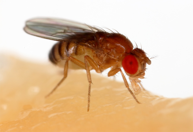
Scientists at the Howard Hughes Medical Institute in Ashburn, Virginia, have used a pair of high-speed electron microscopes to produce a high-resolution digital snapshot of the adult fruit fly brain. Their work – involving 7,062 brain slices and 21 million images – means that researchers can now trace the path of any one neuron to any other neuron throughout the insect's brain. The breakthrough is reported this week in the peer-reviewed journal Cell. "The entire fly brain has never been imaged before at this resolution that lets you see connections between neurons," says Davi Bock, group leader at the Institute's Janelia Research Campus. "Any time you look at images with higher resolution and more completeness, you're going to discover new things." His team's data offers a new tool for researchers studying brain circuitry – the precise webs of neuronal connections that underpin specific behaviours, memories and other functions. The map has already revealed a new cell type and other surprises. The brain of a common fruit fly (or Drosophila melanogaster) is about the size of a poppy seed and contains 100,000 neurons. Each neuron branches into a "starburst" of fine wires that touch the wires of other neurons. Neurons talk to one another through these touchpoints, or synapses, forming a dense mesh of communication circuits. Despite having a million times fewer neurons than a human (which typically has 86 billion), fruit flies are surprisingly sophisticated, explains Bock: "They can learn and remember. They know which places are safe and dangerous. They have elaborate sequences of courtship and grooming."
Scientists can view these wires and synapses with an imaging technique called serial section transmission electron microscopy. First, they infuse the fly's brain with a cocktail of heavy metals. These pack into cell membranes and synapses, eventually marking the outlines of each neuron and its connections. Then the researchers hit slices of the brain with a beam of electrons, which passes through everything except the metal-loaded parts. The resulting images expose the brain's once-hidden nooks and crannies. "It's the same way that x-rays go through your body except where they hit bone," explains Bock. Electron microscopy has, historically, been a slow process. A decade ago, collecting the millions of images necessary for viewing the entire fly brain would have been out of the question, Bock says: "Imagine taking 21 million pictures with your iPhone. You'd be sitting and clicking for decades." He and a group of scientists developed new tools to speed up the process. The team used high-speed cameras and two custom-built systems to rapidly move tissue samples in eight-micrometre increments, allowing them to quickly capture images of neighbouring areas. They were able to image an entire brain slice in less than seven minutes – five times faster than the previous high-throughput camera array, TEMCA1. Bock and his colleagues also benefitted from a custom robotic loader, built at Janelia, able to pick up and move samples automatically. This can be seen in the video at the start of this blog. Creating such cutting-edge technology was no small feat. The work required the collaboration of dozens of Janelia neuroscientists, mechanical engineers and software developers, as well as consultants and scientists at Johns Hopkins University and the MRC Laboratory of Molecular Biology. After a long informal collaboration between these scientists, a formal Janelia project team co-managed by Bock and Janelia software engineer Khaled Khairy was created in 2016 to push the effort to completion. Without the entire team, Bock says, the study wouldn't have been possible: "This is an 'only-at-Janelia' kind of enterprise."
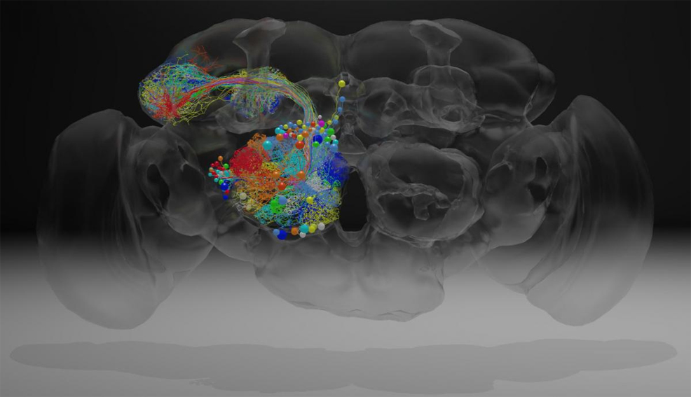
The millions of images collected and stitched together provide an in-depth look at the fly brain – and the chance to explore uncharted areas. Bock's team traced the paths of neurons that reach out to the mushroom body, a region involved in memory and learning. These cells, "olfactory projection neurons", send messages to neurons called Kenyon cells. These cells, in turn, talk to different sets of neurons. Until now, scientists hadn't identified Kenyon cells' conversation partners in a region of the mushroom body called the calyx. Bock's team pinpointed some of these neurons, as well as a previously unknown brain-spanning neuron that also relays information to Kenyon cells. The olfactory projection neurons also appeared to be more tightly bundled together than scientists had thought, Bock says. This bundling suggests an orderly structure in something once believed to be largely random. A better understanding of this circuitry could give scientists insight into fly behaviour. "We think it will tell us something about how the animal learns – how it associates odours with a reward or punishment," explains Bock. Now, more than 20 lab groups are digging into the new dataset, tracing neurons and outlining the brain's circuitry. Bock calls the data a resource that's free to be mined by neuroscientists probing the mind of the fly: "It adds another tool to the toolkit we're using to understand this animal." This achievement follows news earlier in the month about a plan to simulate entire regions of the mouse brain on a supercomputer. Given the rate of progress we are seeing, it is surely just a matter of time before a whole human brain can be either photographed, or replicated via software, with extreme levels of detail.
Comments »
If you enjoyed this article, please consider sharing it:
|