9th August 2021 Monkey brain mapped in 3D at micron resolution Chinese scientists have produced a 3D map of an entire rhesus monkey brain, which is 200 times larger than a mouse brain, at a resolution of just 0.001 mm (1 micron).
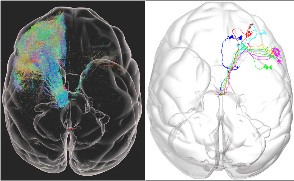
A team from the Chinese Academy of Sciences (CAS) developed a high-throughput technique based on fluorescent imaging, called VISoR. This system works by using an efficient pipeline combining tissue sectioning and clearing, 3D microscopy and semi-automated reconstruction and tracing (SMART). Their study is published in Nature Biotechnology. At present, imaging the entire brain of a mouse – in 3D and micron resolution – can take days. However, high-resolution brain mapping for non-human primates such as monkeys, although highly desired for their potential role in modelling human diseases, has been hindered by a major technical challenge. The brain of a rhesus monkey is simply too big, at more than 200 times the volume of a mouse. To overcome this problem, researchers at the CAS developed Volumetric Imaging with Synchronous on-the-fly-scan and Readout (VISoR). Compared with commonly used 3D optical imaging techniques, VISoR eliminates the time loss caused by moving and pausing while switching fields of view to obtain a series of 2D images, allowing for imaging without blur when the sample is in continuous motion. The result is 3D imaging of large tissue samples at more than 10 times the speed of conventional methods. In addition to the challenge of throughput speed, a further difficulty arises from imaging monkey brains. The cortical folding structures are very complicated and with low tissue transparency. The CAS team solved this by first sectioning the isolated brain into ultra-thin slices (0.3 mm) and then developing reagents to make the samples thoroughly transparent. With their improved VISoR 2.0 system, they completed imaging of a whole macaque brain in 100 hours at a resolution of 1×1×2.5 microns. The total volume of raw image data exceeded 1 petabyte (PB).
Alongside this, the researchers developed efficient algorithms and software to realise automated 3D imaging reconstruction and semi-automated tracking of individual neuronal axon fibres over long distances. Their initial observations revealed "previously unknown characteristics of axonal fibre projection and surprising patterns of fibre turning and routing" in the cortical folds. Prof. David Van Essen, a neuroscientist from Washington University in St Louis, has described this work as a "technical tour de force that marks a stunning advance in our ability to map long-distance connectivity, accurately and efficiently throughout the entire brain of the macaque monkey". He said that besides the technical achievement, their discovery may have profound implications for understanding brain morphogenesis and the principle of "wiring length minimisation". According to the CAS team, VISoR has potential to be extended to other tissues and organs, including samples from clinical pathology. They anticipate that by combining the vast amounts of raw image data with AI analysis, this technique may help to understand the fine 3D structure of the brain and body as well as how they change in various disease conditions, thus facilitating medical diagnostics and drug developments. "Brain connectome at the mesoscopic level is important but so far limited to rodents. This work demonstrates a powerful method that enables researchers to dissect mesoscopic connectome of monkeys at one micron resolution, in four days. It represents a tour de force in this rapidly moving field," said Prof. Wang Xiaojing from New York University. "Hopefully, this technology will be further improved for broader and larger scale applications, to make important contributions to the mapping and understanding of primate and eventually the human brain," said Prof. Duan Shumin from Zhejiang University.
Comments »
If you enjoyed this article, please consider sharing it:
|