9th March 2019 New optical imaging system could spot tiny tumours Researchers at the Massachusetts Institute of Technology (MIT) have demonstrated a new optical imaging system that could enable the discovery of tiny tumours, as small as 200 cells in size, deep within the body.
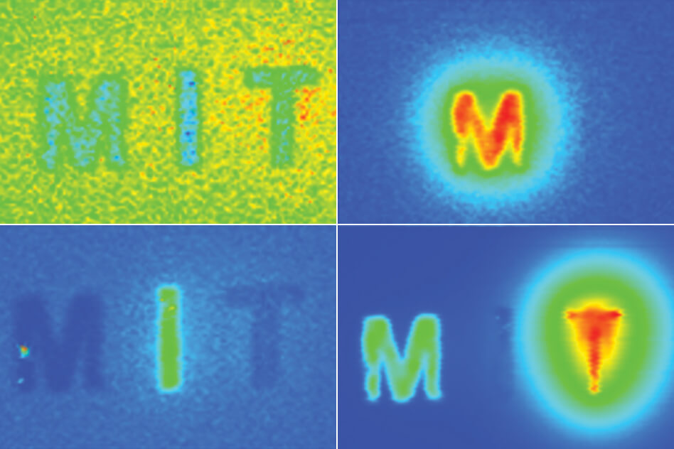
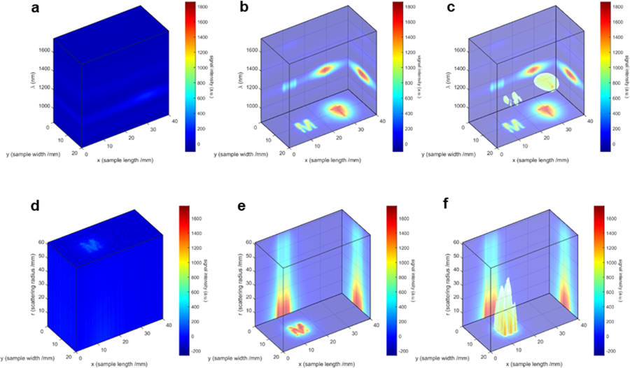
Many types of cancer could be more easily treated if they were detected at an earlier stage. MIT researchers have now developed an imaging system, named "DOLPHIN," which could enable them to find tiny tumours, as small as a couple of hundred cells, among the 37 trillion cells in the entire human body. In a new study, they used their imaging system, which relies on near-infrared light, to track a 0.1-millimetre fluorescent probe through the digestive tract of a living mouse. They also showed that they can detect a signal to a tissue depth of 8 cm, far deeper than any existing biomedical optical imaging technique. It is hoped that this technology could be adapted for early diagnosis of pancreatic and other cancers that are currently difficult to detect until late stages. "We want to be able to find cancer much earlier," says Angela Belcher, Professor of Biological Engineering and Materials Science and the newly-appointed head of MIT's Department of Biological Engineering. "Our goal is to find tiny tumours, and do so in a noninvasive way." Existing methods for imaging tumours all have limitations that prevent them from being useful for early cancer diagnosis. Most have a trade-off between resolution and depth of imaging, and none of the optical imaging techniques can image deeper than about 3 cm into tissue. Commonly used scans such as X-ray computed tomography (CT) and magnetic resonance imaging (MRI) can image through the whole body; however, they can't reliably identify tumours until they reach around 1 cm in size. Belcher's lab set out to develop new optical methods for cancer imaging several years ago. They wanted to create technology that could image very small groups of cells deep within tissue, and do so without any kind of radioactive labelling. Near-infrared light, which has wavelengths of 900-1700 nanometres, is well-suited to tissue imaging, because light with longer wavelengths doesn't scatter as much when it strikes objects, allowing it to penetrate deeper into the tissue. To take advantage of this, the researchers used an approach known as "hyperspectral imaging", which enables simultaneous imaging in multiple wavelengths of light. The new system was tested with a variety of near-infrared, fluorescent, light-emitting probes – i.e. nanoparticles – with various rare-earth elements added through a process called doping. Depending on the choice of element, each of these particles emits near-infrared fluorescent light of different wavelengths. The researchers used algorithms to analyse data from the hyperspectral scans. This helped them identify the sources of fluorescent light of different wavelengths and determine the location of a particular probe. By further analysing light from narrower wavelength bands within the entire near-IR spectrum, it was also possible to determine a probe's depth. The researchers call their system "DOLPHIN", which stands for "Detection of Optically Luminescent Probes using Hyperspectral and diffuse Imaging in Near-infrared."
To demonstrate the potential usefulness of their system, the researchers tracked a 0.1-millimetre-sized cluster of fluorescent nanoparticles that was swallowed and then travelled through the digestive tract of a living mouse. These probes could be modified so that they target and fluorescently label specific cancer cells. "In terms of practical applications, this technique would allow us to non-invasively track a 0.1-millimetre-sized fluorescently-labelled tumour, which is a cluster of only a few hundred cells," said co-author Neelkanth Bardhan, a postdoctoral fellow at MIT. "To our knowledge, no one has been able to do this previously using optical imaging techniques." In ongoing work, they are using a related version of this imaging system to try to detect ovarian tumours at an early stage. Ovarian cancer is usually diagnosed very late, because there is no easy way to detect it when the tumours are still small. They have also begun working on adapting this type of imaging to detect other types of cancer, such as pancreatic and brain cancer, as well as melanoma. "Ovarian cancer is a terrible disease, and it gets diagnosed so late because the symptoms are so nondescript," Belcher says. "We want a way to follow recurrence of the tumours, and eventually a way to find and follow early tumours when they first go down the path to cancer or metastasis. This is one of the first steps along the way in terms of developing this technology." "This is really amazing work," said Guosong Hong, an assistant professor of materials science at Stanford University, who was not involved in this study. "For the first time, fluorescent imaging has approached the penetration depth of CT and MRI, while preserving its naturally high resolution, making it suitable to scan the entire human body."
Comments »
If you enjoyed this article, please consider sharing it:
|