|
|
|
|
|
|
|
7th April 2026
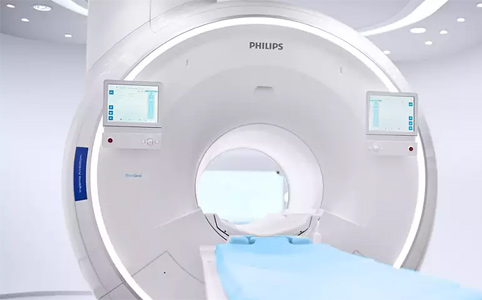
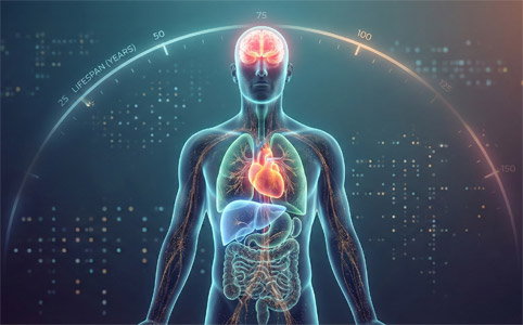
AI cuts MRI scan times by more than half
Artificial intelligence is helping hospitals dramatically reduce MRI scan times while also boosting image quality, with a leading cancer centre in Amsterdam reporting a drop from 23 minutes to just 9 minutes.
Read more... |
|
|
|
|
|
|
25th March 2026
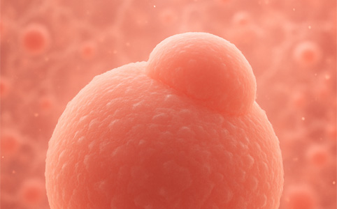
World's first lab-grown oesophagus
In recent years, scientists have made progress growing replacement body parts in the lab, but complex tubular organs remain especially difficult. Now, a team has created the first lab-grown oesophagus, successfully implanted in an animal.
Read more... |
|
|
|
|
|
|
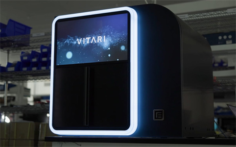
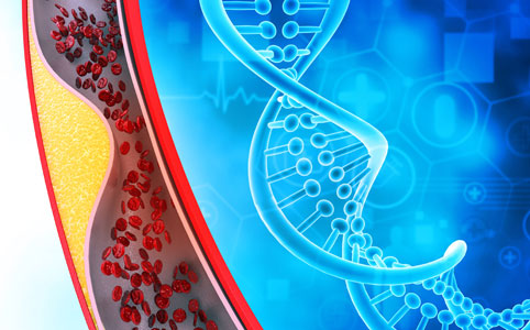
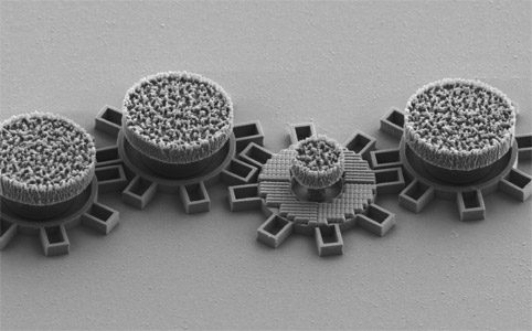
25th February 2026
First benchtop $100 genome system
The long-promised $100 genome may finally be entering mainstream commercial territory. Element Biosciences has launched what it describes as the first benchtop sequencing platform explicitly designed to deliver whole human genomes at that price – a step beyond earlier room-scale demonstrations by rival firms.
Read more... |
|
|
|
|
|
|
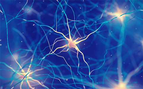
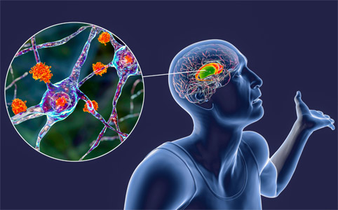
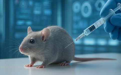
13th February 2026
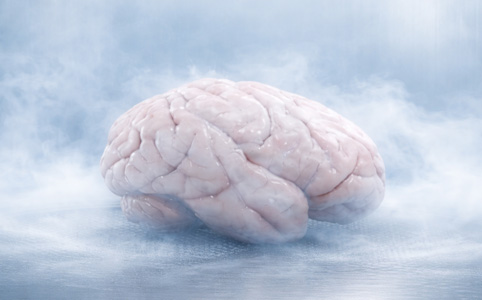
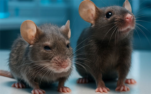
Protein restores aging brain stem cells
Researchers have identified a key protein that restores aging neural stem cells in laboratory models. Their work sheds new light on how the brain's regenerative capacity declines over time.
Read more... |
|
|
|
|
|
|
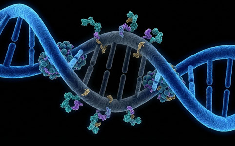
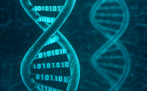
30th September 2025
DNA data storage could arrive within 3–5 years
An international consortium says DNA could soon become a practical medium for digital archives. A new report points to the first use cases emerging in just 3–5 years, with densities up to 500 million times greater than conventional drives.
Read more... |
|